This is the time and platform to raise my voice and to be heard
Title: Spontaneous pneumoperitoneum in neonates
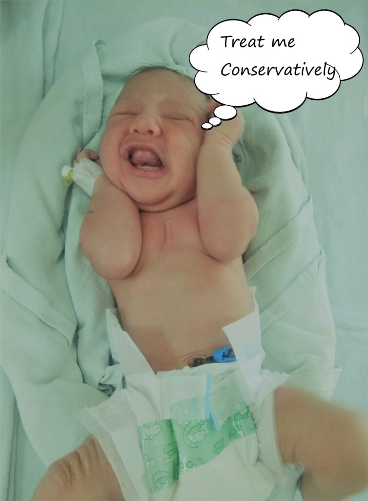
Sometimes, I have an intuition that my treating surgeon could be more observant and wait patiently in some rare situations. He should contemplate a conservative approach rather than performing an invasive surgical procedure on me. As I believe that each scar on me (my abdomen) puts an invisible scar on the heart of my mother. I want my surgeon to be more sensitive towards me. It is said that “a good surgeon knows how to operate, but a best surgeon is the one who knows, when not to operate.”
Here I have explained concisely, this condition known as spontaneous pneumoperitoneum in neonates. Pneumoperitoneum is classically a surgical emergency. However, pneumoperitoneum can rarely present without any gastrointestinal perforation/pathology, especially in neonates. These neonates may improve mainly with conservative management or may require abdominocentesis or abdominal drain placement. Thus, pneumoperitoneum without any gastrointestinal (GI) perforation or peritonitis is entitled spontaneous pneumoperitoneum. The condition is rare in clinical practice and it is seen in approximately 5% of neonates with pneumoperitoneum.

The condition poses a dilemma regarding surgical management of the neonate. But before considering the management, it is paramount to understand the etiopathogenesis of spontaneous pneumoperitoneum. The source of air in spontaneous pneumoperitoneum is air-leak from the lungs as shown in the flowchart. This is more common with high and prolonged airway pressure during mechanical ventilation or CPAP (one-half to two-third cases). It may be preceded or simultaneously seen with pneumothorax or pneumomediastinum.
")
In case of spontaneous pneumoperitoneum, the onset is abrupt usually following mechanical ventilation. Usually there is absence of systemic signs of sepsis. Associated malformations may be present in approximately 50% of patients. An algorithm for the management of neonate with pneumoperitoneum is presented.
")
In any paediatric patient, especially neonate with radiographs showing pneumoperitoneum, a comprehensive clinical evaluation is mandatory. Repeated abdominal examination for signs of peritonitis. Pulmonary signs, especially for presence of pneumothorax and sepsis screen and blood parameters (including CBC) must be carried out to supplement the diagnosis. In presence of sepsis or marked abdominal distension and suspicious cases, ultrasound with Doppler is mandatory for ruling out perforation/visceral pathology.
Abdominocentesis under ultrasound guidance and/or abdominal drain placement should be contemplated to rule out perforation and to direct further management. Finally, knowledge and experience with this condition would greatly impact the management and planning of pediatric patients, particularly neonates with pneumoperitoneum. This would prevent unnecessary surgery leading to undue morbidity and mortality. Remember, you have to treat me and not my X-ray.
")

Further reading:
- Gupta R, Bihari Sharma S, Golash P, Yadav R, Gandhi D. Pneumoperitoneum in the newborn: is surgical intervention always indicated? J Neonatal Surg. 2014;3(3):32.
- Gupta R. Spontaneous Pneumoperitoneum in Paediatric Patients: Dilemmas in management. J Indian Assoc Pediatr Surg. 2018;23:115-22.
- Duan SX, Sun ZB, Wang GH, Zhong J, Ou WH, Fu MX, et al. Diagnosis and treatment of pediatric benign pneumoperitoneum: A case report series of 9 patients. Medicine. 2017; 96:e5814.
- Gupta R. Spontaneous pneumoperitoneum: Discerning from radiological imaging. J Neonatal Surg. 2021;10:6
Dr. Rahul Gupta
MS, M.Ch. Paediatric Surgery FMAS
Associate Professor, Department of Paediatric Surgery
SMS Medical College, Jaipur, Rajasthan, India
email: meetsurgeon007@gmail.com